Cryo-electron Tomography

cryo-et-description

Cryo-ET

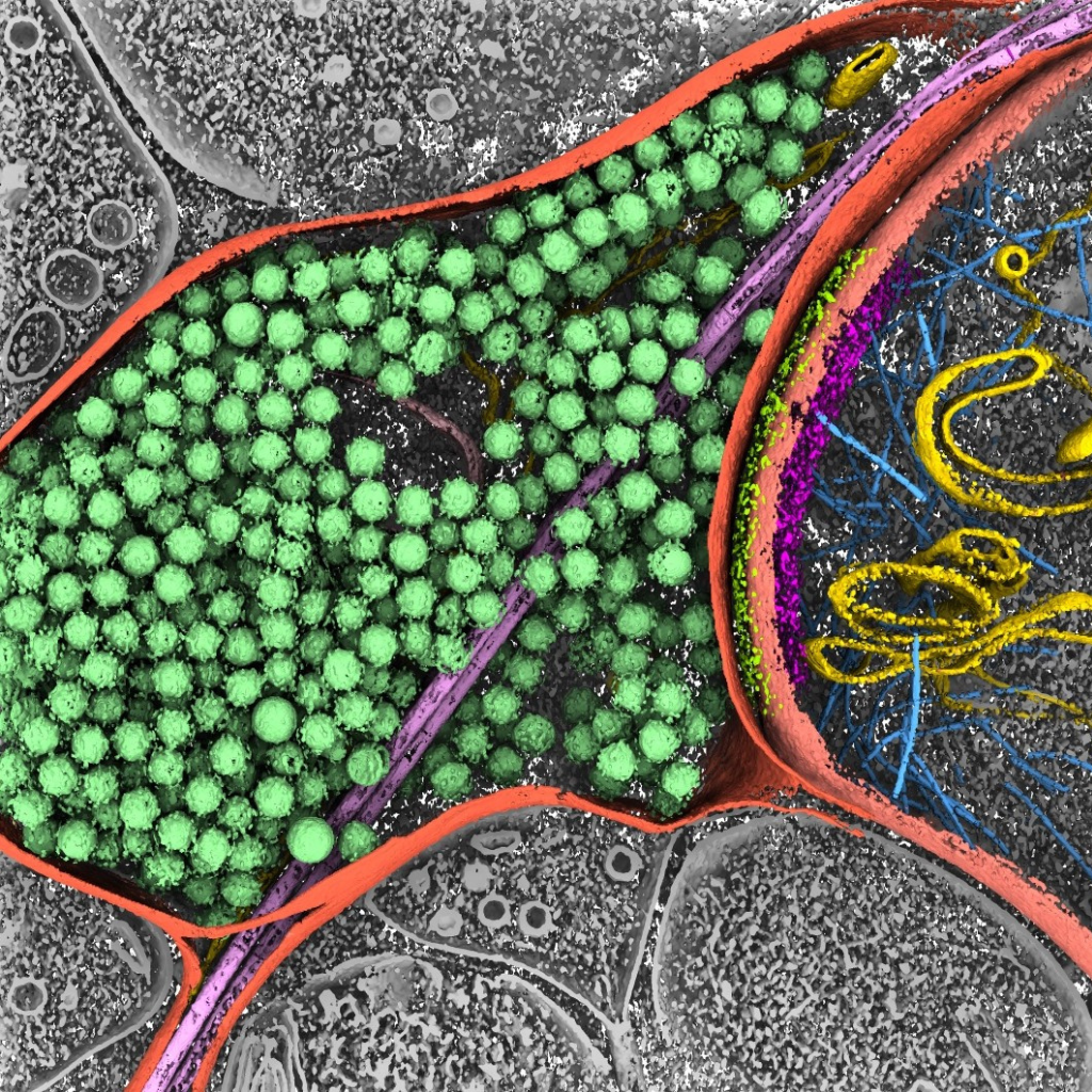
Cryo-electron tomography (Cryo-ET) involves acquiring 2D electron projections at various sample tilt angles and using these projections to generate a 3D image of the sample with nanometer-scale resolution. This process is similar to medical imaging techniques like CT scans.

Cryo-ET allows for in-situ three-dimensional structural analysis of particles with non-uniform morphological distributions ranging from nanometers to sub-micrometers.

By analyzing the in-situ three-dimensional structure of samples, Cryo-ET can reveal information regarding the distribution of proteins on drug delivery carriers, the arrangement of drugs inside these carriers, the hierarchical structure of particles, and interactions between particles.

Cryo ET

Application Scenarios

Cryo-electron tomography is widely used in the study of fundamental life processes, enabling high-resolution resolution of various cellular ultrastructures such as organelles, cell membranes, and protein complexes, contributing to a deeper understanding of cellular structure and function. This technology plays a crucial role in investigating the structure and lifecycle mechanisms of pathogens (viruses and bacteria), aiding in revealing the mechanisms of disease occurrence and transmission pathways, thereby providing a basis for drug discovery and treatment. Cryo-electron tomography can present the structures of proteins, complexes, and ligands in near-natural states, offering essential information for the design, optimization, and screening of structure-based antibody drugs, enzymes, and small molecule drugs, thereby advancing drug development.

Talk to Us

Thank you for your support of Nanomega CryoAI. We look forward to providing you with excellent structural analysis services. If you have any questions or needs, please feel free to contact us.